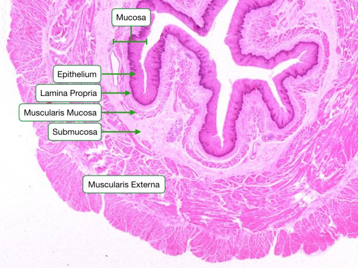
GI tract histology model labeled, a powerful tool in medical research and clinical practice, provides a detailed representation of the gastrointestinal tract’s structure and function. This model, meticulously labeled with specific markers, offers unparalleled insights into the intricate workings of the GI system, aiding in the diagnosis, treatment, and prevention of a wide range of gastrointestinal disorders.
The labeled histology of the GI tract unveils the distinct layers and components of this vital organ system, revealing the cellular and molecular mechanisms underlying its functions. It serves as a cornerstone for understanding the normal physiology of the GI tract, as well as the pathological changes associated with various diseases.
GI Tract Histology Model
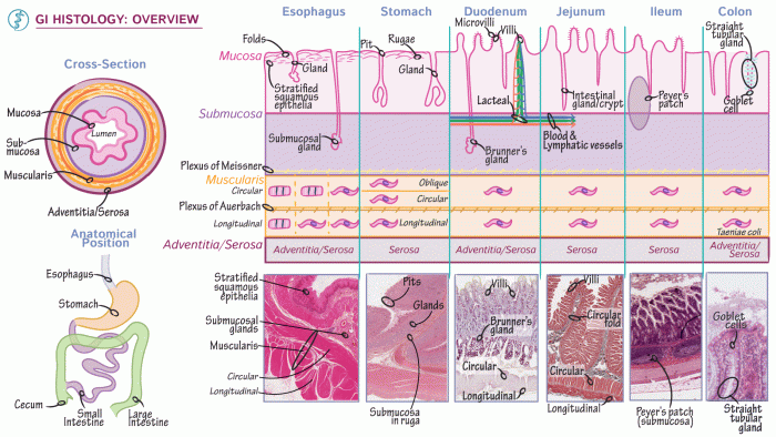
The gastrointestinal (GI) tract histology model is a valuable tool for studying the structure and function of the digestive system. It provides a comprehensive representation of the different components and layers of the GI tract, allowing researchers to visualize and analyze the tissue in detail.
The GI tract histology model is typically created using a combination of techniques, including tissue fixation, sectioning, and staining. The resulting model can be used for a variety of purposes, including:
- Studying the normal structure of the GI tract
- Identifying and characterizing pathological changes
- Developing new diagnostic and therapeutic approaches
Components of the GI Tract Histology Model
The GI tract histology model consists of the following components:
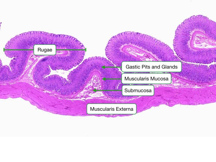
- Mucosa:The mucosa is the innermost layer of the GI tract and is responsible for absorption and secretion. It is composed of a layer of epithelial cells, a lamina propria, and a muscularis mucosae.
- Submucosa:The submucosa is a layer of connective tissue that lies beneath the mucosa. It contains blood vessels, nerves, and lymphatic vessels.
- Muscularis externa:The muscularis externa is a layer of smooth muscle that is responsible for propelling food through the GI tract. It is composed of an inner circular layer and an outer longitudinal layer.
- Serosa:The serosa is the outermost layer of the GI tract and is composed of a layer of mesothelium and a layer of connective tissue.
Labeled Histology
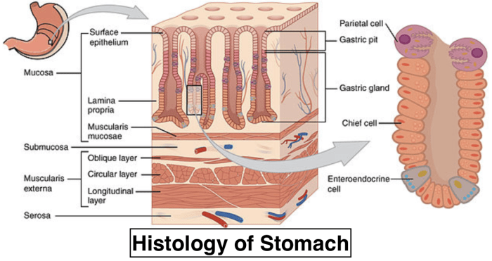
Labeled histology is a specialized technique used in histology, the study of tissues, to enhance the visualization and identification of specific structures within a tissue sample. In GI tract histology, labeled histology plays a crucial role in understanding the structure and function of the gastrointestinal tract.
Labeled histology involves the application of specific labels or markers to tissue sections. These labels can be antibodies, dyes, or other substances that bind to particular molecules or structures within the tissue. Once labeled, the tissue sections are examined under a microscope, allowing researchers and diagnosticians to visualize and analyze the distribution and localization of specific proteins, molecules, or cellular components.
Various Labeling Techniques
There are various labeling techniques used in GI tract histology, each with its advantages and disadvantages. Some commonly used techniques include:
- Immunohistochemistry (IHC): IHC uses antibodies to bind to specific proteins within the tissue. This technique is commonly used to identify and localize specific cell types, growth factors, or other proteins of interest.
- In situ hybridization (ISH): ISH uses labeled probes to bind to specific nucleic acid sequences within the tissue. This technique is used to visualize the expression of specific genes or to detect the presence of specific pathogens.
- Histochemical staining: Histochemical staining uses dyes or other chemicals to react with specific molecules or structures within the tissue. This technique is commonly used to visualize the distribution of mucins, lipids, or other components of the extracellular matrix.
Benefits and Limitations
Labeled histology offers several benefits for research and diagnosis in GI tract histology:
- Enhanced visualization: Labeled histology allows for the visualization of specific structures or molecules within the tissue, providing a more detailed understanding of the tissue’s composition and organization.
- Identification of specific cell types or molecules: Labeled histology enables the identification and localization of specific cell types, growth factors, or other molecules of interest, providing insights into the cellular and molecular mechanisms underlying GI tract function and disease.
- Diagnosis of diseases: Labeled histology is a valuable tool in the diagnosis of various GI tract diseases, such as cancer, inflammatory bowel disease, and infectious diseases. By identifying specific markers or patterns of expression, labeled histology can aid in the accurate diagnosis and classification of these diseases.
However, labeled histology also has some limitations:
- Potential for false positives and false negatives: The accuracy of labeled histology depends on the specificity and sensitivity of the labeling technique used. False positives or false negatives can occur due to non-specific binding or cross-reactivity.
- Limited information on tissue function: Labeled histology provides information on the localization and expression of specific molecules or structures but does not provide direct information on the functional state of the tissue.
- Time-consuming and expensive: Labeled histology can be a time-consuming and expensive technique, especially for large or complex tissue samples.
Applications of the GI Tract Histology Model: Gi Tract Histology Model Labeled
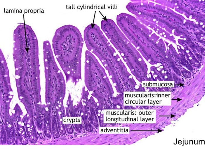
The GI tract histology model is a valuable tool for research and clinical practice, providing insights into the structure and function of the GI tract.
In research, the model is used to study GI diseases, drug development, and surgical planning. It enables researchers to examine the microscopic features of the GI tract and identify changes associated with various conditions.
Studying GI Diseases
The GI tract histology model allows researchers to study the histopathological changes associated with GI diseases such as inflammatory bowel disease, celiac disease, and colorectal cancer.
- By examining tissue samples under a microscope, researchers can identify characteristic histological features that aid in diagnosing and classifying GI diseases.
- The model also helps in studying the progression of GI diseases and evaluating the efficacy of different treatment strategies.
Drug Development
The GI tract histology model is used in drug development to assess the effects of new drugs on the GI tract.
- Animal models are often used to study the histological changes induced by drug candidates, helping researchers evaluate potential side effects and interactions with the GI tract.
- The model also aids in optimizing drug delivery systems and dosage regimens to ensure optimal absorption and efficacy.
Surgical Planning, Gi tract histology model labeled
In surgical planning, the GI tract histology model provides valuable information for surgeons.
- By examining tissue samples, surgeons can determine the extent of disease involvement and plan appropriate surgical interventions.
- The model helps identify anatomical landmarks and variations, minimizing the risk of complications during surgery.
Future Directions and Advancements
The field of GI tract histology modeling is rapidly evolving, with new technologies and advancements emerging all the time. These advancements have the potential to further improve the accuracy and utility of the GI tract histology model, making it an even more valuable tool for research and clinical practice.
One of the most promising areas of research is the use of artificial intelligence (AI) and machine learning (ML) to analyze labeled histology data. AI and ML algorithms can be used to identify patterns and relationships in data that are not easily detectable by the human eye.
This can help to improve the accuracy of diagnosis and prognosis, and it can also lead to the discovery of new biomarkers for GI diseases.
Potential of Artificial Intelligence and Machine Learning
- Improved accuracy of diagnosis and prognosis
- Discovery of new biomarkers for GI diseases
- Development of personalized treatment plans
Another area of research that is likely to have a significant impact on the GI tract histology model is the development of new imaging techniques. These techniques can provide more detailed and accurate images of the GI tract, which can help to improve the diagnosis and treatment of GI diseases.
New Imaging Techniques
- Confocal laser endomicroscopy (CLE)
- Optical coherence tomography (OCT)
- Magnetic resonance imaging (MRI)
Finally, there is a need for further research to develop new methods for preparing and staining GI tissue samples. These methods can help to improve the quality of the images obtained, which can lead to more accurate diagnosis and prognosis.
Methods for Preparing and Staining GI Tissue Samples
- Immunohistochemistry
- In situ hybridization
- Electron microscopy
Popular Questions
What is the purpose of a GI tract histology model labeled?
A GI tract histology model labeled serves as a detailed representation of the gastrointestinal tract’s structure and function, providing insights into the normal physiology and pathological changes associated with GI diseases.
How is labeled histology used in GI tract research?
Labeled histology enables the visualization and identification of specific cell types, proteins, and other molecules within the GI tract, facilitating the study of cellular and molecular mechanisms underlying GI function and disease.
What are the benefits of using a GI tract histology model labeled?
The GI tract histology model labeled offers a comprehensive view of the GI system, aiding in the diagnosis and classification of GI diseases, guiding treatment decisions, and assessing treatment outcomes.
